Hip Joint Featured Image

Hip Joint – Bones, ligaments, blood supply and innervation |Anatomy|

We are going to be looking at the hip joint, its articulations, movements, blood supply, and innervation.

So, the hip joint is a ball and socket synovial joint, forming the connection between the lower limb and the pelvic girdle.

And the hip joint is also multi-axial meaning that it rotates on more than one axis. Which allows for a wider range of movement. Of course, the hip joint is designed for stability and weight-bearing.

Hip joint

The head of the femur articulates with the lunate surface of the acetabulum of the pelvis.

Head of femur

The lunate surface is concave, which is like the shape of a crescent moon as the name lunate suggests. And the hip joint can also be referred to as the acetabulofemoral joint.

lunate surface


Both the lunate surface of the acetabulum and the head of the femur are covered by hyaline cartilage. And hyaline cartilage is simple cartilage found on many articulating surfaces.

So, the acetabulum is the part of the pelvis where the ilium, ischium, and pubis bones merge and its concave almost entirely encompasses the head of the femur. Which contributes to the stability of the joint.

Hip-Bones-Pelvic-Girdle

At the center of the acetabulum is a non-articulating surface which is known as the acetabular fossa. This part of the acetabulum contains loose connective tissue.

acetabular fossa


The acetabular labrum is the fibrocartilaginous collar or lip that surrounds the bony rim of the acetabulum and it bridges across the acetabular notch.

acetabular notch


The labrum of the acetabulum increases the stability of the hip joint by deepening the acetabulum and increasing the area of articulation with the head of the femur.

labrum of the acetabulum


The ball of the ball and socket hip joint is the rounded head of the femur. Which sits within the concavity of the acetabulum, which is our socket of the joint.

The head of the femur is entirely covered by hyaline cartilage except for an area called fovea capitis femoris.
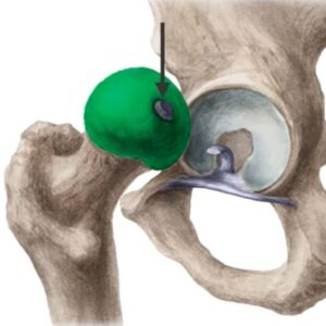
fovea capitis femoris


This is where the ligament of the head of the femur connects the femur at the fovea to the acetabular fossa, the transverse acetabular ligament, and the margins of the acetabular notch.

margins of the acetabular notch


 Capsule OF the Hip Joint:

So, the joint capsule of the hip joint attaches to the acetabular labrum and the transverse acetabular ligament proximally but is underneath the fibrous capsule.

underneath the fibrous capsule

The joint capsule is a strong fibrous capsule that can accommodate a wide range of movements and the capsule is strengthened and reinforced by the help of three ligaments.

Anteriorly and superiorly by the iliofemoral ligament, which connects the anterior and inferior iliac spine and the acetabular rim to the femoral intertrochanteric line. The iliofemoral ligament is the strongest of the hip joint ligaments.

iliofemoral ligament


Inferiorly and anteriorly, the pubofemoral ligaments arise from the obturator crest and the superior ramus of the pubis and blend with the capsule and the medial part of the iliofemoral ligament.

ischiofemoral ligament


Posteriorly, the ischiofemoral ligament seen here from a posterior view connects the ischial part of the acetabular rim of the neck of the femur.

The fibers from the three ligaments are arranged in a spiral fashion around the hip joint which helps stabilize the joint by pulling the head of the femur medially into the acetabulum.

This reduces the amount of muscle energy required to maintain a standing position and prevents the hyperextension of the hip as well as excessive abduction.


Movements of the Hip Joint:

So the range of hip movement in the hip joint include

  • Flexion – Movement of the leg forward
  • Extension – Movement of the leg backward
  • Abduction – Movement of the leg laterally
  • Adduction – Movement of the leg medially towards the midline of the body
  • Medial rotation – Internal rotation of the thigh towards the midline.
  • Lateral rotation – Outward rotation of the thigh from the midline of the body.
  • Circumduction – Conical precise 360degree movement of the leg.

Hip Movement


Blood Supply of the Hip Joint:

Blood is supplied to the hip joint primarily by the medial and lateral circumflex femoral arteries – the medial shown on the left and the lateral shown on the right. Both of these arise from the deep femoral artery.

deep femoral artery

And also by the artery to the head of the femur which runs within the ligament of the head of the femur which is shown in green and the artery to the head of the femur is a branch of the obturator artery.

obturator artery


Innervation of the Hip Joint:

(Green color indicates nerve)

1. Innervation of the hip joint comes anteriorly from the femoral nerve. 

anteriorly from the femoral nerve

2. Inferiorly from an articular branch of the anterior division of the obturator nerve.

anterior division of the obturator nerve

3. Poster superiorly from the superior gluteal nerve.superior gluteal nerve
4. Laterally from the articular branch of the sciatic nerve.
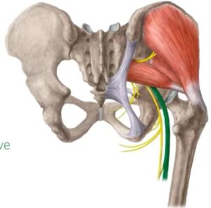
sciatic nerve.


For further advice do reach out to your local doctor or family doctor.

Do share this blog with your friends and family!

Read More
Scapula Anatomy featured Image (Final)

Anatomy and Function Of The Scapula – Human Anatomy


Let’s talk about the scapula and answer the question what is the scapula? What are its primary bony landmarks?

What is Scapula?

The Scapula also called the shoulder blade lies on the posterior part of the body. The scapula is a flat triangular bone placed in the thoracic cage (human rib cage).

Extending from the level of the second rib to the seventh rib. It provides support to the muscles of the forelimb. Articulation for the humerus (upper arm bone) at the glenoid cavity and it is joined to the clavicle in front.

The scapula or shoulder blade is a major bony component of the shoulder and functions to connect the upper extremity with the trunk of the body.

The scapula articulates with two other bones:

  1. Laterally with the humerus at the glenoid cavity.
  2. Superiorly with the clavicle at the acromial process.


Side Determination of Scapula:

How do we determine the side of the scapula? Basically, the spinous process should always face backward. The glenoid cavity should face laterally that is away from the body.

The lateral border is always thick while the medial border is thin.


Anatomical Features of the Scapula:


Surfaces:

The costal surface is concave and it faces the thoracic cage. It has three longitudinal ridges and gives attachment to the intermuscular septa. There is another thick ridge adjoining the lateral border. This part of the bone is almost rod-like. It acts like a lever for the action of the serratus anterior in the overhead abduction of the arm.

The dorsal surface gives attachment to the spine of the scapula which divides the surface into a smaller supraspinous fossa and a larger infraspinous fossa. The two fossa are connected to each other by the spinal glenoid notch. It is situated between the spine of the scapula and the glenoid angle.

Angle: 

The angle includes the superior angle, inferior angle, and lateral angle.

  • The superior angle is covered by the trapezius.
  • The inferior angle is covered by the latissimus dorsi and it lies just opposite to the superior angle it moves forwards around the chest. When the arm is abducted.
  • The lateral angle or the glenoid angle faces laterally and away from the body.


Borders:

  • The medial border is the longest and faces the vertebral column. It’s on the medial side of the bone, so it’s called the medial border of the scapula. It’s also the border that is closest to the vertical column, so it’s also called a “vertebral border”.
  • The lateral border is the border of the scapula facing the humerus. It’s also close to your armpit, so it’s called “axillary border” as well.
  • The superior border, which is the shortest and thinnest.


Bony Features of the Scapula:

The acromion also articulates with the clavicle and so between the acromion and clavicle there is a joint it’s called the acromioclavicular (AC) joint. It’s a synovial plane joint. The clavicle articulates with the scapula. It allows you to move your arm up and down and the clavicle and scapula label to move your arm so it gets mobility.

Acromioclavicular (AC) joint


The spine of the scapula has the trapezius muscle that is anchors to give support to the scapula.

Spine of the scapula


The fossa is a shallow concave surface, so this fossa is on the back of the scapula and above the spine of the scapula so it’s called a supraspinous fossa.

supraspinous fossa

The infraspinous fossa is the fossa below the spine of the scapula.

Infraspinous fossa

The glenoid fossa of the scapula or the glenoid cavity is a part of the shoulder. It is shallow, which is located on the anterior side of the scapula.

Glenoid cavity fossa

Supraglenoid tubercle has found small projection found at the superior margin of the glenoid cavity. The supraglenoid tubercle is the origin point of the long head of the biceps.


Supraglenoid tubercle (bicep)

On the inferior margin of the glenoid cavity, you will find the infraglenoid tubercle. This is also an important landmark to remember since the long head of the triceps arises from the structure.

Infraglenoid tubercle

This coracoid process is important for muscle attachment, in fact, three muscles are the pectoralis minor muscle, the short head of the biceps muscle, and the coracobrachialis muscle all have attachments to the coracoid process.

Coracoid process

On the suprascapular notch, we have a ligament called the suprascapular ligament that goes across. So, you have the suprascapular artery that goes to the ligament.

Suprascapular notch


If you have any questions about Scapula Anatomy please feel free and leave a comment.

Do share this blog with your friends and family!

Read More
Knee Featured Image

Anatomy Of The Knee

For the anatomy of the knee joint, we’ll begin by looking at an anterior view of the right knee.
The knee joint is made up of three bones femur, patella, and tibia. There are also medial condyle and lateral condyle. The patella articulates with the femur and the patella is the biggest sesamoid bone in the body.

There is a small amount of fibula which does not make up the knee joint. Rather it articulates with the tibia (Shinbone), which does make up the knee joint.

The tibia has a tibial tuberosity which can get inflamed when it gets inflamed it is known as Osgood-Schlatter disease (OSD).

anterior knee view

The knee joint is special because there between the joints is a fibrocartilaginous structure called the meniscus. There is medial and lateral meniscus and these menisci are basically shock absorbers and help stabilize join.

It also has a role in the distribution of the synovial fluid. The knee joint is further stabilized by many ligaments the lateral collateral ligament and the medial collateral ligament.

The quadriceps tendon essentially attaches and overlies the patella bone and then forms the patella ligaments. The patella bone has a patella ligament that runs and attaches to the tibial tuberosity which can get inflamed.

The patella ligament is actually the continuation of quadriceps tendons formed by the rectus femoris muscle and the vastus muscles of the thigh.

knee

The side view of the knee joint and introduce the bursa, the bursa is fluid-filled sacs that help reduce friction. So, the femur, tibia, and the patella.

The meniscus is the fibrocartilaginous structure that sits between the joints. The Quadriceps tendon comes down over and basically encapsulates the patella in front, forming the patella ligament.

Sometimes the patella ligament is also known as the patella tendon and essentially onto the tibial tuberosity.

Side view of the knee

The bursa can get inflamed because of irritation between the skin and bone from overuse and friction. The pre patella bursa can get inflamed this is known as housemaid’s knees. This is because supposedly house-maid are on their knees and scrub the floor.

This will cause friction between the skin and the patella leading to inflammation of the bursa between the two.

The knee joint is the largest joint in the human body. The function of the knee joint is to allow movement of the leg.

The popliteal artery is a continuation of the femoral artery. This artery supplies blood to the knee joint through numerous small branches.

Inside the knees, you will find the smooth articular cartilage that covers the joint surface.

Symptoms:

Some of the associated symptoms for knee pain are,

  • Swelling
  • Instability
  • Weakness
  • Stiffness


Causes:

The incongruent surface is the biggest reason why the knee is more prone to develop injury and pay. However, are the reasons which one can experience knee pain is due to malalignment or poor tracking of patella bone. Due to tightness and weakness of the thigh muscles, due to degenerative changes in the joint also called arthritis of the joint.

Due to repeated injury and sometimes due to some systemic diseases.


For further advice do reach out to your local doctor or family doctor.

Do share this blog with your friends and family!


Read More
Spine Featured Image

Vertebral Column Anatomy

Your spine is also called your backbone or vertebral column is composed of 33 bones called vertebrae. Which provides your body with support and protects your spinal cord from injury.

The vertebrae can be divided into five group’s cervical, thoracic, lumbar, sacral, coccyx. Each of which has unique features suited to its functions there are

  • 7 Cervical vertebrae numbered C1 to C7.
  • 12 Thoracic vertebrae numbered T1to T12.
  • 5 Lumbar vertebrae numbered L1 to L5.
  • 5 Sacral vertebrae numbered S1 to S5.
  • Coccyx

The upper 24 vertebrae are articulating separated by intervertebral discs the sacral vertebra and the bones of the coccyx are fused.
The cervical vertebrae (neck) which are the vertebrae of your neck support your head. Which weighs around 10 to 13 pounds.
C1 and C2 are specialized Vertebrae,

  • Allow for the greatest range of motion of all the vertebrae
  • C1 is called “ATLAS”. Ring Shaped and attaches directly to your skull
  • C1 allows you to nod your head
  • C2 is called “AXIS”. Serves as an axis around which C1 pivots
  • C1 is able to pivot on C2 thanks to a specialized process called DENS, or ODONTOID process

The Thoracic vertebrae are the vertebrae of your mid and upper back. Hold the ribcage and protect your heart and lungs. They have a limited range of motion.

The Lumbar vertebrae are the vertebra of the lower back. Bear the weight of your upper body and are larger in order to support the stress. Especially when you lift something heavy.

The Sacral vertebrae connect your spine to your hip bones these vertebrae are fused together with your hip bones, they form the pelvic girdle.

The coccyx or tailbone is made up of four fused which provide an attachment point for ligaments and muscles of the pelvic floor.

Parts of a Vertebra:

A vertebra has three parts

  1. Body
  2. Vertebral arch
  3. Processes

Spine 2

The body bears weight, the vertebral arch houses, and the spinal cord and the processes allow for muscle attachment. The vertebral arch is made up of two supporting pedicles and two laminae.

Space inside which houses the spinal cord is called the vertebral foramen. Under each pedicle spinal nerves exit and pass through the intervertebral foramina.

Seven bony processors arise from each vertebral arch to form facet joints and processes for muscle attachment.

Facet joints allow for back motion each vertebra has two pairs of facet joints one pair connects to the vertebra above and one pair that connects to the vertebra below.

There is also a pair of transverse processes and a spinous process.

Between the vertebras are the intervertebral discs, which provide cushioning and prevent the bones from rubbing together.

Discs are composed of an annulus. Which is made up of several layers or lamina of fibrocartilage.

Criss-crossing fibrous bands attach between the bodies of the vertebrae above & below and a gel-filled center called the nucleus.

The nucleus distributes the pressure evenly within each disc during compression.

The nucleus is full of proteoglycans large molecules with sugar subunits that are very hydrophilic. This structure swells with water unless it experiences constraints from the surrounding tissues.

spine 3

Hence, the nucleus absorbs fluid at night or when you are lying down and then this fluid is pushed out during the days. When you are upright and pressure is applied to the intervertebral discs. We shorten as we age because the discs lose the ability to reabsorb fluid during rest and they flatten and become more brittle.


If you have any questions about Vertebral Column please feel free and leave a comment.

Do share this blog with your friends and family!


Read More
Bone Cancer Signs (Featured Image)

10 Signs Of Bone Cancer


Bone cancer is a malignant form of cancer that begins in the bone and destroys normal bone cells. This unusual kind of cancer affects both adults and children.

Most people who have a disease in their bones develop it from the secondary spread of another cancer. This condition is called Metastasis.

Some of the common risk factors associated with the development of bone cancer previous chemotherapy, diseases such as hereditary retinoblastoma, tuberous sclerosis, or prior treatment with radiation therapy.

Keep reading to learn more about the symptoms of bone cancer.


1. Bone Pain:

One of the most common complaints of patients with bone cancer is consistent pain in the affected bone. The pain may not be constant at first, it can come and go but gradually becomes chronic.

Typically, the pain is worse at night. However, it can also ache during the day, such as pain in the legs while walking.

As cancer spreads the pain tends to remain there all the time. With more physical activity, it increases more.


2. Swelling and Redness:

You will experience swelling and tenderness in the affected area if you develop bone cancer. However, inflammation may not be visible in the early stages of cancer.

It usually shows up after a few weeks. You may fee a lump or mass deposition on the site of the tumor. If a bump develops in the bones of the neck, then inflammation can occur in the back of the throat. This condition will lead to problems swallowing.


3. Fractures Due Bone Weakness:

Abnormal growth of cells weakens the bone as the disease grows. In most cases, the bones do not fracture or break.

However, you might complain of sudden severe pain in your limb. This sign is especially notable after it has been tender for a few months. Bone cancer can make the bones weak that they can break even after a minor fall.


4. Limping:

Limping is a common symptom seen majorly in people who develop bone cancer in the leg. It is a condition where a person is not able to walk correctly.


5. Unexplained Weight Loss:

Sudden loss of weight that is not caused by stress, physical workouts, or a changed diet plan can be a sign of bone cancer.

Typically, you will experience weight loss along with bone pain. This state requires immediate medical attention. As the body won’t be able to get rid of wastes easily, you will experience constipation.

A loss of appetite will follow, leading to unintended weight loss.


6. Fever:

Among the set of different complications associated with bone cancer, contracting a low-grade fever is a telling sign. Though not every time, but in a lot of cases, bone cancer can trigger a fever. Not only will you have a temperature, but other symptoms as well.

The temperature is usually low-grade, from mild to moderate. It should be around 99 degrees but can be higher in some cases.


7. Anemia:

If you have been running short of breath and feeling tired for a few days now, there is a possibility that you are anemic. Anemia is a telling sign of cancer, including bone cancer.

It occurs with the scarcity of red blood cells in the body. As the bones weaken, the body produces white blood cells to combat the abnormal cells causing cancer in the body, which reduces the red blood cells count.

Mild anemia may not be as bothersome as moderate anemia, which can cause headaches and weakness.


8. Loss Of Appetite:

You might experience weight loss as the body remains exhausted all the time. When the cancerous tumor starts spreading in the body, you might lose your willingness to eat.

Food might begin tasting bad or lose its flavor. As the cancerous cells in the body suck all nutrients from the meal, it leaves you drained out and disinterested in food.


9. Disturbed Sleep:

Trouble in sleeping or disturbed sleep is another symptom of bone cancer. This symptom occurs because of episodes of pain in the bones. You can have difficulty falling asleep.

You might also wake up multiple times in the night. Once you wake up in the middle of the night, you might lay awake.


10. Fatigue:

As with other types of cancer, bone cancer leads to fatigue and weakness. This symptom can certainly impact your quality of life. Physical and mental fatigue with any underlying cause can be a sign of a condition like cancer. You may have persistent tiredness for a long period of time.


Diagnoses

Bone cancer can be preliminary diagnose with the analysis of bone X-ray or MRI image. An X-ray image provides a low-priced diagnostic tool for diagnosis and visualization of bone cancer as compared to MRI.


We recommend you to take your doctor’s advice for proper guidance and discuss these symptoms with your doctor.

Do share this blog with your friends and family!

Read More
fibula Featured Image

Fibula: Feature, Fracture Types, and Symptoms


The ankle is made up of three bones the tibia or shin bone, the fibula or calf bone, and the talus. The most common ankle fracture usually involves the fibula.

Side Determination of Fibula:


How do we determine the side of a fibula? The upper end of the head is slightly expanded in all directions. The lower end or lateral malleolus is expanded anteroposterior and flattened from side to side.


fibula lower end

On the medial side of the lower end, there is a triangular articular facet anteriorly and a deep malleolar fossa facing posteriorly.


Features of Fibula:


The Fibula has an upper end, a shaft, and a lower end.

Fibula upper.end

Now let’s look at the upper end of the fibula. The upper end is slightly expanded in all directions.

The fibula is the cylindrical lateral bone of the leg that is very small and thin. If you look at the placement of this bone which is parallel to the tibia. The tibia is the main part with the fibula acting as a pin that has to be inserted in the loop.

It is also called the calf bone because it’s present at the back of the leg. The fibula bone it’s not weight-bearing. The tibia is the weight-bearing bone of the leg.

The fibula is so non-weight bearing that it can be taken out of it and use if for bone reconstruction elsewhere in the body.

So its main functions are is forming the ankle joint. And a site of muscle attached as we have got lots of muscles attached to the fibula and the tibia and the interosseous membrane. So muscle attachments forming the ankle joint.

Basically, fibula fractures are very common, and fibular fractures commonly happen due to minor trauma.

The fibula is the lateral and smaller bone of the leg. It is very thin as compared to the tibia.


What are the types of fibula fractures?


Fibula fractures occur around the knee, ankle, and middle of the leg. There are types of fracture, which can also affect recovery and treatment. These types include:

  1. Avulsion fracture – a fracture in which a small part of the bone gets pulled off.
  2. Lateral malleolus fracture – a break around the ankle.
  3. Fibular head fracture – a break near the knee.
  4. Shaft fracture – a break that often affects the middle of the leg due to direct impact.
  5. Stress fracture – a hairline fracture due to repetitive injury e.g. Soccer, rugby, and snowboarding.


What are the symptoms of a fracture?


Other than pain and swelling, other signs of a fibula fracture include:

• bruising and tenderness
• deformity in the lower part of the leg
• pain that gets worse when putting pressure on the leg
• tingling or numbness, which usually happens if there is a neurovascular injury

Other joints and bones involved, such as the tibia.


Educational fibula fracture platting procedure:


First, the surgeon will make an incision over the fracture and a clamp (surgical tool) is used to hold the broken bone in place using special instruments a hole is drilled in the bone and a screw is used to hold the fracture in the correct position.

Next, the distal fibula plate is placed onto the bone and held with two pins then using special instruments screws are inserted through the plate to hold it onto the bone. After the ankle fracture has been fixed with a plate. Your surgeon drills holes in both the fibula and the tibia just above the ankle joint. The implant is inserted through the holes until it reaches the other side. Then your surgeon pulls back on the handle positioning the button against the bone.

Finally, the round button is placed against the plate and your surgeon pulls on each suture (Thread-like material) until the desired tension is achieved. This completes the ankle fracture repair.


We recommend you to take your doctor’s advice for proper guidance.

Do share this blog with your friends and family!


Read More
clavicle fracture featured image

Clavicle Fracture: Symptoms, Causes and Recovery/Rehabilitation

Most clavicle fractures will heal without an operation. They quite often managed in either a sling or brace to try to help support the weight of the arm to prevent the fracture from displacing and this also provides pain relief.

You have sustained a fracture to the middle portion of your collarbone also known as the clavicle.

Clavicle Fracture


As the above picture demonstrates that there is a common injury and it will heal itself naturally with the passage of time this normally, takes approximately six weeks but pain and swelling can be ongoing for three to six months because of the nature of the injury. There may always be a lump in this area it should however not be painful and have no long-term effects.

fracture sling


You will have been provided with a sling. This should be worn for the first two weeks. Only it should be removed regularly to perform the exercises.

You may need to take painkillers or anti-inflammatories, especially in the early stages. Some of these injuries require a follow-up appointment with an upper limb specialist. Certain injury types can lead to delayed healing.

  • One of the things that can slow down healing is smoking. If you do smoking it would be advisable to stop at least for the duration of the healing process.
  • You can start driving again when you can comfortably control the car. And obviously, you can’t drive if you’re still in the sling.
  • You can return to work when you feel comfortable and sports can be resumed at six weeks. Keep in mind though that pain and swelling can be ongoing. When you start impact activities.


Symptoms


  • Pain that increases with shoulder movement
  • Swelling
  • Tenderness
  • Bruising


Causes


  • Falls- Such as falling onto your shoulder or onto your outstretched hand.
  • Sports injuries- Such as a direct blow to your shoulder on the field, rink, or court.
  • Vehicle trauma- From a car, motorcycle, or bike accident.


Initial Exercises to be taken as part of your rehabilitation:


It’s really important in the first stage of your rehabilitation to remove your sling to do the following exercises three to four times a day.

  1.  Hand and Wrist Exercise: You can move your wrist in upwards and downwards position getting a good stretch of that joint. You can use a pair of socks that you make a ball and hold and squeeze to add some resistance to this exercise. This is good to help to reduce the swelling.
  2.  Elbow Exercise: You need to practice straightening your elbow and bending it towards your mouth. Elbow also works in rotation and moves your hand from facing palm up to palm down.
  3.  Rollback Shoulder Exercise: You should bring your shoulders back so that, you get a stretch across the front of your chest. You should hold this position for approximately 30 seconds and repeat this four to five times a day.
  4.  Pendulum exercises: Hold on to a firm surface bring your feet into a step starts position and gently lean forward letting your arm come forward with it. You can then move your arm in forwarding and backward momentum. Just like the pendulum from a clock and also do with sideways movement, clockwise rotation. Do this exercise for a minute or a minute and a half again four to five times a day.


These exercises are only to be undertaken after 3 weeks:


The next stage of your rehabilitation is about gently starting to move your shoulder. This exercise is known as an active-assisted range of movement.

The first movement direction is to hold on to your hand with your good arm and gently guide it up in the air. You should repeat this 10 times nice and slowly and thinking about the quality of the movement.

The second movement is a rotational way for this get a stick and place your arm firmly with an overhand grip onto the stick. Use your arm in a sideways outer movement. Keep your elbow close by your side.

The exercise you should do about 10 repetitions on both exercises about 4 to 5 times a day.


These exercises are only to be undertaken after 6 weeks:


The third stage of rehabilitation is about regaining the range of movement and strength in your shoulder.

The first exercise is bringing your arms straight up in front of you as far as you can do comfortably. It’s important to focus on your posture while doing it.

The second exercise is to your thumb outwards and do a similar movement but in a big arc. Once again just go as far as you feel able to and each day hopefully this will improve a little bit.


We recommend you to take your doctor’s advice for proper guidance.

Do share this blog with your friends and family!


Read More
Osteoporosis Featured Image

Osteoporosis – causes, symptoms, diagnosis and treatment


Osteo refers to bones and porosis means pores. So, osteoporosis is when there’s a higher breakdown of bone. A decrease in bone density to the point of potential fracture.

Looking at a cross-section of a bone, there’s a hard-external layer known as the cortical bone and a softer internal layer of spongy bone that is composed of trabeculae. The trabeculae are like a framework of beams that give structural support to the spongy bone. The cortical bone, in turn, is made up of many functional, pipe-like units called osteons, which run through the length of the bone.

In the center of these osteons, there are hollow spaces called Haversian canals, which contain the blood supply and innervation for the bone cells. Around the Haversian canals, there are concentric lamellae, which look a bit like tree rings.

The lamellae have an organic part, which is mostly collagen, and an inorganic part called hydroxyapatite, which is mostly calcium phosphate. In between neighboring lamellae, there are spaces called lacunae, which contain bone cells called osteocytes.

Osteoporosis

At first glance, the bone may appear motionless and unchanging, but it’s actually a very dynamic tissue.

Bone Remodeling:

In fact,

  • Spongy bone is replaced every 3 to 4 years.
  • Compact bone is replaced every 10 years.

In a process called bone remodeling which has two steps:

  1. Bone resorption: when specialized cells called osteoclasts break down bone.
  2. Bone formation: which is when another type of cells called osteoblasts form new bone.


Bone remodeling as a whole is highly dependent on serum calcium levels, which, in turn, are kept in the normal range by a balance between parathyroid hormone (PTH), Calcitonin, and Vitamin D.

Parathyroid hormone is produced by the parathyroid glands in response to low serum calcium, and it increases bone resorption to release calcium into the bloodstream.

On the other hand, calcitonin is produced by the thyroid gland in the response to high serum calcium, so it opposes the action of PTH- therefore promoting bone formation and decreasing bone resorption.

Finally, vitamin D promotes calcium absorption in the gut, so it increases serum calcium, promoting bone formation and decreasing bone resorption.

Peak Bone Mass

The balance between their regulatory factors results in a peak bone mass, usually by age 20 to 29 and this usually occurs earlier in females than in males. Factors that determine the peak bone mass are genetics. For example, people of African descent tend to have greater bone mass and nutrition.

Finally, strength training increases peak bone mass, as well as hormones like estrogens and androgens that inhibit bone resorption.

When osteoclasts break down bone faster than the osteoblasts can rebuild, it results in the lowering of the bone mass and eventually in osteoporosis.

Causes

Spongy bone and cortical bone, these bone changes increase the risk of fracture, and they are known as fragility or pathologic fractures. Some bones like the vertebrae, shoulder blades, and ribs consist mainly of spongy bone, so they are at great risk of fragility fractures.

Factors that accelerate bone mass loss and increase the risk of osteoporosis are low estrogen levels, like after menopause, and low serum calcium.

Additional factors include alcohol consumption, smoking, drugs that decrease calcium absorption from the gut through antagonism of vitamin D, and drugs like heparin and L-thyroxine.

Another factor is physical inactivity, as seen in astronauts in a zero-gravity environment where they just don’t use their musculoskeletal system as hard as when they’re on earth. As a result, bone deposition decreases due to a lack of stress, while resorption increases.

There are also diseases that can cause osteoporosis like

  • Turner syndrome
  • Hyperprolactinemia
  • Klinefelter syndrome
  • Cushing syndrome
  • Diabetes mellitus.


Now, the two most common types of osteoporosis are

1. Postmenopausal osteoporosis

In postmenopausal osteoporosis, decreased estrogen levels lead to increased bone resorption.

2. Senile osteoporosis

People with senile osteoporosis don’t usually have symptoms until a fracture occurs.

Symptoms

The most common type of fractures are vertebral fractures, also known as compression fractures, and it occurs when one or more bones in the spine weaken and shatter. Vertebral fractures cause back pain, height loss, and a hunched posture.

Femoral neck fractures and distal radius fractures can also occur, and they’re often associated with postmenopausal osteoporosis.

Diagnosis

Osteoporosis is usually diagnosed with a dual-energy X-ray absorptiometry or DEXA scan which tests for bone density.

Treatment  

Treatment for osteoporosis usually relies on bisphosphonate drugs like Alendronate and Risedronate. If osteoporosis is really advanced, teriparatide, a recombinant parathyroid hormone can be used.


We recommend you to take your doctor’s advice for proper guidance.

Do share this blog with your friends and family!


Read More