The tibia, commonly known as the shinbone, is one of the most important bones in the human body. Located between the ankle and the knee, it carries most of our body’s weight and plays a crucial role in walking, running, and everyday movement. Because of its load-bearing function, the tibia is also one of the bones most prone to fractures.

ANATOMY OF THE TIBIA:

The tibia is the larger of the two bones in the leg, positioned between the knee and the ankle. It has a hollow canal filled with bone marrow and a strong outer layer known as the cortex, which provides strength.

Fisioterapia Sportiva Milano - Dott. Creperio Fisioterapista

◼️Tibial plateau – top part of the tibia near the knee
◼️Tibial plafond – bottom part of the bone near the ankle.
◼️Tibial shaft – the central part where most of the fractures occur.

WHAT IS THE TIBIAL SHAFT?

The tibial shaft is the middle portion of the tibia, lying between the flared ends just below the knee (tibial plateau) and just above the ankle (tibial plafond). Unlike the ends, which are broader and flatter, the shaft is more triangular in shape.

SIGNS OF TIBIAL SHAFT FRACTURES:

Tibia / Fibula Fracture Specialist | 🩺Singapore Sports & Orthopaedic  Surgeon

Fractures of the tibial shaft typically occur with substantial traumatic injuries. Common signs of these fractures include:

◼️Sharp pain in the shin.
◼️Visible deformity of the leg.
◼️Bruising and swelling around the shin.
◼️Inability to bear weight on the leg.

Because such injuries often occur with other trauma, doctors usually examine the entire body to rule out
hidden injuries.

For more information, talk to a healthcare provider.

If you have any questions about TIBIAL SHAFT FRACTURES, Please feel free to leave a comment.

Do share this blog with your friends and family!

Read More

WHAT IS A HERNIATED DISC?

Your spine is made of bones (vertebrae) stacked on each other. Between them are discs that act like cushions. Each disc has a tough outer layer and a soft inner part (nucleus). When the disc gets damaged and the inner material pushes out, it’s called a herniated or slipped disc. This can press on nerves, causing pain and other issues.

SYMPTOMS:

What is Slip Disc? Symptoms of Slip Disc

◼️Muscle Weakness: Nerve compression reduces strength and function.
◼️Difficulty in Walking: poor coordination or pain while moving.
◼️Burning or Aching: Indicates nerve irritation.
◼️Numbness: Loss of sensation in compressed nerve areas.
◼️Standing or Sitting: Pain worsens when standing or sitting.

CAUSES:

◼️Poor Posture: Sitting or standing the wrong way strains the spine.
◼️Smoking: Reduces blood supply, causing discs to wear out faster.
◼️Too Much Sitting or Wrong Lifting: Long sitting or lifting heavy things incorrectly hurts the discs.
◼️Being Overweight: Extra body weight puts more pressure on the spine.
◼️Getting Older: Discs naturally weaken and wear down with age.

TYPES:

Spine Disorders back pain concept with degenerative disc or bulging and Herniated or thinning discs and osteophyte formation on a skeletal vertebra as a close up of a vertabrate.

◼️Normal Disc: Healthy spine with no issues.
◼️Degenerative Disc: Natural wear and tear over time, reducing flexibility.
◼️Bulging Disc: Disc bulges out, may press on nerves, causing discomfort.
◼️Herniated Disc: Outer layer of disc ruptures, inner material may leak.
◼️Thinning Disc: Disc loses height,
reducing spinal support.

Extra changes: Bone spurs (osteophytes) and endplate hardening can also happen, causing stiffness.

For more information, talk to a healthcare provider.

If you have any questions about  WHAT IS A HERNIATED DISC? Please feel free to leave a comment.

Do share this blog with your friends and family!
Read More
aging

How Aging Affects Your Musculoskeletal System and What You Can Do About It :

As we age, our bodies undergo a series of changes, and the musculoskeletal system is no exception. The musculoskeletal system, which includes bones, muscles, tendons, ligaments, and joints, plays a crucial role in maintaining our mobility, strength, and overall quality of life. Understanding how aging affects this system and taking proactive steps to address these changes can help you maintain your independence and well-being as you grow older.


aging

How Aging Affects the Musculoskeletal System:

1. Bone Density Loss:
  • Osteoporosis: One of the most significant age-related changes is the loss of bone density, leading to osteoporosis. Bones become thinner and more brittle, increasing the risk of fractures and breaks.
  • Bone Remodeling: With age, the process of bone remodeling slows down, meaning that bone resorption (breaking down) outpaces bone formation. This imbalance contributes to reduced bone mass and strength.
2. Muscle Mass and Strength Decline:
    • Sarcopenia: Age-related muscle loss, known as sarcopenia, begins around the age of 30 and accelerates after the age of 60. This condition results in decreased muscle mass and strength, impacting balance and mobility.
    • Muscle Fiber Changes: The number of muscle fibers decreases with age, and the remaining fibers tend to become larger but less efficient, leading to reduced muscle function and endurance.
    3. Joint Changes:
    • Cartilage Degeneration: Cartilage, the smooth tissue that cushions joints wears down over time. This can lead to osteoarthritis, characterized by joint pain, stiffness, and decreased range of motion.
    • Joint Flexibility: The synovial fluid that lubricates joints decreases with age, leading to stiffer and less flexible joints.
    4. Tendon and Ligament Changes:
      • Tendon and Ligament Stiffness: Tendons and ligaments become stiffer and less elastic with age, reducing joint stability and increasing the risk of injuries like sprains and strains.
      • Tendon Repair Slows Down: The ability of tendons and ligaments to repair and heal slows with age, making recovery from injuries more prolonged.
      5. Postural Changes:
        • Kyphosis: The natural curvature of the spine may increase with age, leading to a condition known as kyphosis, where the upper back becomes excessively rounded. This can affect posture and balance.
        • Height Loss: Compression of the spinal discs and changes in bone density can lead to a gradual loss of height over time.


        aging

        What You Can Do About It:

        Maintaining the health of your musculoskeletal system as you age involves a combination of lifestyle choices, exercise, and medical care. Here’s how you can address and mitigate the effects of aging on your musculoskeletal system:

        1. Engage in Regular Exercise:
        • Weight-Bearing Exercises: Activities like walking, jogging, and dancing help stimulate bone formation and maintain bone density.
        • Strength Training: Resistance exercises, such as lifting weights or using resistance bands, can help combat muscle loss, improve muscle strength, and enhance overall physical function.
        • Flexibility and Balance Exercises: Incorporate exercises like yoga, stretching, and tai chi to improve joint flexibility, balance, and coordination, reducing the risk of falls.
        2. Prioritize a Nutrient-Rich Diet:
        • Calcium and Vitamin D: Ensure adequate intake of calcium and vitamin D to support bone health. Foods like dairy products, leafy greens, fortified cereals, and fatty fish are excellent sources.
        • Protein: A sufficient protein intake is crucial for muscle maintenance and repair. Include lean meats, beans, nuts, and dairy products in your diet.
        • Anti-Inflammatory Foods: Incorporate foods with anti-inflammatory properties, such as fruits, vegetables, nuts, and fatty fish, to support joint health and reduce inflammation.
        3. Maintain a Healthy Weight:
        • Avoid Excessive Strain: Maintaining a healthy weight reduces the stress on your joints, particularly the knees, hips, and lower back. Excess weight can exacerbate joint pain and increase the risk of osteoarthritis.
        • Balanced Diet and Exercise: Combine a balanced diet with regular exercise to achieve and maintain a healthy weight.
        4. Address Joint Health:
        • Manage Arthritis Symptoms: If you have arthritis, work with your healthcare provider to manage symptoms through medication, physical therapy, and lifestyle changes.
        • Protect Your Joints: Use supportive devices like braces or orthotic inserts if recommended by your doctor, and avoid activities that put excessive stress on your joints.
        5. Monitor Bone Health:
        • Bone Density Testing: If you’re at risk for osteoporosis, discuss bone density testing with your healthcare provider to assess bone health and take preventive measures if needed.
        • Regular Check-Ups: Keep up with regular medical check-ups to monitor bone health and address any issues promptly.
        6. Avoid Harmful Habits:
        • Quit Smoking: Smoking accelerates bone loss and impairs the healing process. Quitting smoking is crucial for maintaining bone health.
        • Limit Alcohol Consumption: Excessive alcohol intake can interfere with bone health and increase the risk of fractures. Limit alcohol consumption to moderate levels.
        7. Focus on Fall Prevention: 
        • Home Modifications: Make your home safer by removing tripping hazards, installing grab bars in the bathroom, and ensuring adequate lighting.
        • Fall Prevention Exercises: Incorporate exercises that improve balance and coordination to reduce the risk of falls and related injuries.


        For more information talk to a healthcare provider.

        If you have any questions about  How Aging Affects Your Musculoskeletal System, please feel free to leave a comment.

        Do share this blog with your friends and family!

          Read More
          JOINTS SURGERY

          Joint’s Replacement Surgery: What You Need to Know

          Joint replacement surgery is a common and highly effective procedure for relieving pain, improving mobility, and enhancing the quality of life for individuals with severe joint damage. Whether due to arthritis, injury, or other conditions, when conservative treatments like medication and physical therapy no longer provide relief, joint replacement may be the next step. Whether you’re considering this surgery for yourself or a loved one, understanding what to expect before, during, and after the procedure can help you make informed decisions and prepare for a successful recovery.

          joint pain

          What is Joint Replacement Surgery?

          Joint replacement surgery involves removing the damaged or diseased parts of a joint and replacing them with artificial components, called prostheses. These prostheses are typically made of metal, plastic, or ceramic materials designed to mimic the natural movement of a healthy joint. The goal of the surgery is to relieve pain and restore function to the joint, allowing patients to return to their normal activities with improved mobility and less discomfort.

          The most common types of joint replacement surgeries include:

          1. Knee Replacement – Replace the knee joint with an artificial knee, commonly used for osteoarthritis or other knee conditions.

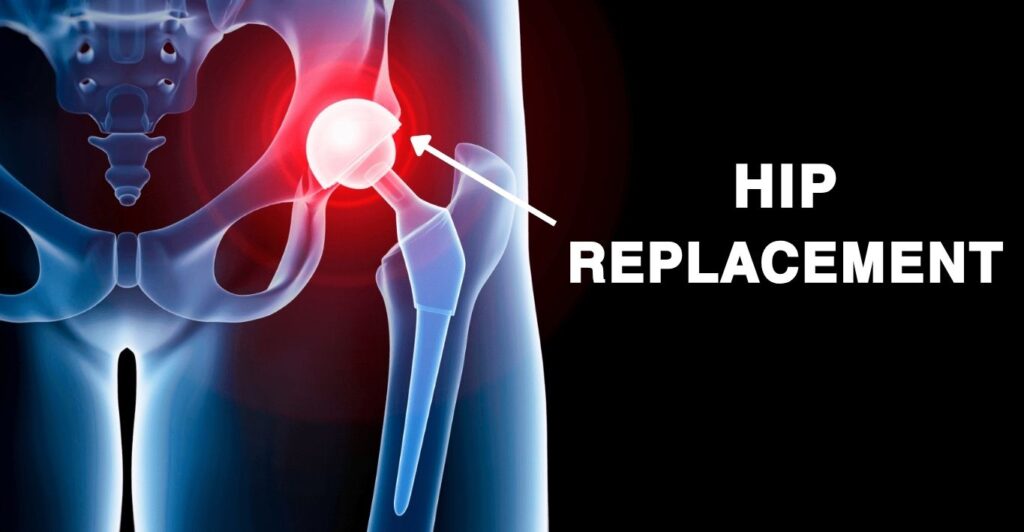
          2. Hip Replacement – Replace the hip joint with an artificial hip, often used to treat arthritis or severe fractures.

          3. Shoulder Replacement – Replace the shoulder joint with an artificial shoulder, typically for severe arthritis or rotator cuff injuries.

          4. Ankle Replacement – Replace the Ankle joint with an artificial Ankle, commonly used for total ankle arthroplasty.

          Each type of joint replacement is tailored to address specific conditions, such as osteoarthritis, rheumatoid arthritis, post-traumatic arthritis, or other degenerative joint diseases.

          joint pain

          When is Joint Replacement Surgery Recommended?

          Joint replacement surgery is typically recommended for individuals with severe joint damage that significantly impacts their quality of life. Joint replacement surgery is considered when joint pain and dysfunction severely impact a person and other treatment options have been exhausted.

          The most common conditions that lead to joint replacement include:

          1. Osteoarthritis – The most common reason for joint replacement, osteoarthritis is a degenerative joint disease that causes the breakdown of cartilage, leading to pain, stiffness, and swelling.

          2. Rheumatoid Arthritis – This autoimmune disorder causes inflammation in the joints, leading to pain and deformity. Over time, it can severely damage the joint, making replacement necessary.

          3. Post-Traumatic Arthritis – This type of arthritis develops after an injury to the joint, such as a fracture or dislocation, which can lead to long-term damage and degeneration.

          4. Avascular Necrosis – A condition where the blood supply to the bone is disrupted, leading to bone death and joint collapse, often necessitating joint replacement.

          5. Severe Joint Injuries – In some cases, severe fractures or joint injuries that cannot be repaired with other treatments may require joint replacement.

          Joint replacement is usually considered when:

          1. Chronic Joint Pain – Pain persists despite non-surgical treatments like physical therapy, medications, or lifestyle changes.

          2. Limited Mobility – Difficulty performing daily activities, such as walking, climbing stairs, or getting in and out of chairs, due to joint stiffness and pain.

          3. Reduced Quality of Life – The joint pain and mobility limitations significantly impact your ability to enjoy life, affecting your mental and emotional well-being.

          4. Joint Deformity – Noticeable deformity of the joint, often due to advanced arthritis, which cannot be corrected by other means.

          5. Severe Pain – Persistent pain that limits daily activities and isn’t relieved by non-surgical treatments such as medications, physical therapy, or injections.

          6. Loss of Function – Significant loss of joint function that affects the ability to perform routine activities like walking, climbing stairs, or grasping objects.

          7. Advanced Joint Damage – Extensive damage to the joint due to conditions such as osteoarthritis, rheumatoid arthritis, or traumatic injury.

          Your orthopedic surgeon will evaluate your condition through a combination of physical examinations, imaging tests (like X-rays or MRIs), and a review of your medical history before recommending surgery.

          Recovery and Rehabilitation:

          Recovery from joint replacement surgery is a gradual process that involves:

          1. Physical Therapy – Physical therapy is essential to restore movement and strength in the new joint. You’ll likely start with simple exercises in the hospital and continue with outpatient therapy after discharge.

          2. Pain Management – Pain and swelling are normal after surgery, but they can be managed with prescribed medications and ice packs.

          3. Mobility Aids – You may need to use crutches, a walker, or a cane for several weeks to months after surgery to assist with walking.

          4. Follow-Up Appointments – Regular follow-up visits with your surgeon will monitor your progress and address any concerns. These appointments are crucial to ensuring proper healing and the long-term success of the joint replacement.

          5. Returning to Activities – While most people can return to normal activities within a few months, it’s important to avoid high-impact activities that could damage the new joint. Low-impact exercises, like swimming, cycling, and walking, are encouraged.


          For more information talk to a healthcare provider.

          If you have any questions about  Joint Replacement Surgery, please feel free and leave a comment.

          Do share this blog with your friends and family!
            Read More
            Deformity Correction

            Understanding Deformity Correction: Techniques and Advances

            Deformity correction is a specialized area of orthopedic medicine focused on realigning bones and joints to their proper anatomical position. Whether due to congenital conditions, trauma, disease, or growth disturbances, deformities can significantly impact an individual’s quality of life, causing pain, functional limitations, and aesthetic concerns. This blog aims to provide a comprehensive overview of deformity correction, exploring the techniques, processes, and advancements in this critical field.

            What Is Deformity Correction?

            Deformity correction involves the surgical or non-surgical realignment of bones and joints to restore normal function and appearance. The primary goal is to improve the patient’s mobility, reduce pain, and enhance overall quality of life. This process can address a wide range of conditions, including:

            • Congenital deformities (e.g., clubfoot, scoliosis)
            • Post-traumatic deformities (e.g., malunited fractures)
            • Degenerative diseases (e.g., arthritis)
            • Growth disturbances (e.g., limb length discrepancies)

            Techniques In Deformity Correction :

            1. Acute Deformity Correction :


            Acute Deformity Correction

            Acute deformity correction is a medical procedure aimed at rapidly realigning bones or joints to their proper anatomical position. This procedure is often employed in the treatment of fractures, dislocations, and congenital or acquired deformities. The process involves several steps and considerations, which can be categorized into preoperative planning, surgical techniques, and postoperative care.

            Preoperative Planning:

            1. Assessment and Diagnosis:

            • Imaging Studies: X-rays, CT scans, and MRIs are used to assess the extent and nature of the deformity.
            • Physical Examination: A thorough physical examination helps to understand the functional impact of the deformity.
            • Patient History: Medical history, including previous treatments and surgeries, is reviewed.

            2. Planning:

            • Correction Strategy: Deciding whether to use internal fixation (e.g., plates, screws) or external fixation (e.g., external fixators).
            • Simulation: Computer-assisted simulations or models may be used to plan the correction precisely.
            • Patient-Specific Considerations: Age, overall health, and bone quality are considered to tailor the approach.

            Surgical Techniques:

            1. Anesthesia:

            • General or regional anesthesia is administered based on the patient’s condition and the procedure’s complexity.

            2. Exposure and Preparation:

            • Incision: A surgical incision is made to access the affected bone or joint.
            • Soft Tissue Management: Careful handling of soft tissues to minimize damage and facilitate healing.

            3. Osteotomy:

            • Bone Cutting: An osteotomy (surgical cutting of bone) is performed to correct the deformity.
            • Realignment: The bone segments are realigned to the desired position.

            4. Fixation:

            • Internal Fixation: Plates, screws, or rods are used to hold the bone in the corrected position.
            • External Fixation: External fixators are applied to stabilize the bone from outside the body.

            5. Adjustment and Verification:

            • Intraoperative Imaging: X-rays or fluoroscopy are used to verify the correction.
            • Adjustments: Fine adjustments are made as needed to ensure proper alignment.

            Postoperative Care:

            1. Recovery:

            • Monitoring: Regular monitoring for signs of infection, proper healing, and correct alignment.
            • Pain Management: Medications and other strategies are used to manage postoperative pain.

            2. Rehabilitation:

            • Physical Therapy: Initiated to restore function, strength, and range of motion.
            • Weight-Bearing: Gradual progression in weight-bearing activities based on healing progress.

            3. Follow-Up:

            • Regular Check-Ups: Scheduled visits to monitor healing and detect any complications early.
            • Imaging Studies: Periodic X-rays or other imaging to assess bone healing and alignment.

            Risks and Complications:

            • Infection: Risk of infection at the surgical site.
            • Nonunion or Malunion: Failure of the bone to heal properly or in the correct position.
            • Hardware Issues: Problems with internal or external fixation devices.
            • Nerve or Vessel Injury: Potential damage to surrounding nerves or blood vessels.

            Acute deformity correction requires a multidisciplinary approach involving orthopedic surgeons, radiologists, anesthesiologists, and rehabilitation specialists to ensure optimal outcomes. The success of the procedure largely depends on meticulous planning, precise surgical execution, and comprehensive postoperative care.


            2. Gradual Deformity Correction :


            Gradual Deformity Correction


            Gradual deformity correction is a technique used to slowly and precisely realign bones or joints over time. This approach is typically employed for more complex deformities, including those resulting from congenital conditions, trauma, or disease processes like osteomyelitis or growth disturbances. The method relies on the body’s natural healing processes and usually involves the use of external fixation devices. Here’s an overview of how gradual deformity correction works:

            Preoperative Planning :

            1. Assessment and Diagnosis:

            • Imaging Studies: X-rays, CT scans, and MRI are used to evaluate the deformity in detail.
            • Physical Examination: A thorough examination assesses the functional impact and the extent of the deformity.
            • Patient History: Reviewing medical history, previous treatments, and any coexisting medical conditions.

            2. Planning:

            • Correction Strategy: Deciding the method and timeline for correction. Computer-assisted planning and simulations are often used.
            • Device Selection: Choosing the appropriate external fixator device, which can be monolateral, circular (Ilizarov), or hexapod-based systems.

            Surgical Technique:

            1. Anesthesia:

            • General or regional anesthesia is administered to the patient.

            2. Surgical Procedure:

            • Incisions and Exposure: Small incisions are made to place the fixator pins or wires.
            • Osteotomy: A controlled cut (osteotomy) is made in the bone at the site of deformity to enable gradual movement.

            3. Application of External Fixator:

            • Pin or Wire Insertion: Pins or wires are inserted into the bone segments through the skin and are attached to the external frame.
            • Frame Assembly: The external fixator frame is assembled around the limb, connecting the pins or wires, allowing for precise control of bone movement.

            Gradual Correction Process:

            1. Distraction Phase:

            • Initiation: Typically starts a few days after surgery to allow initial healing. The process is known as distraction osteogenesis.
            • Adjustments: Gradual adjustments are made to the fixator (usually daily), moving the bone segments very slowly (approximately 1 mm per day) to correct the deformity.
            • Monitoring: Regular follow-ups and imaging (X-rays) to ensure proper alignment and to adjust the correction as needed.

            2. Consolidation Phase:

            • Bone Healing: Once the desired correction is achieved, the bone is allowed to consolidate (harden and heal) in its new position. The fixator remains in place during this phase.
            • Reduced Adjustments: Adjustments are minimized or stopped, and the bone is monitored for proper healing and strength development.

            Postoperative Care and Rehabilitation:

            1. Recovery:

            • Monitoring: Close monitoring for signs of infection, proper alignment, and complications.
            • Pain Management: Appropriate pain management strategies are employed.

            2. Physical Therapy:

            • Early Mobilization: Encouraged to maintain joint mobility and muscle strength, tailored to the patient’s tolerance and progress.
            • Weight-Bearing: Gradual progression of weight-bearing activities as allowed by the surgeon.

            3. Follow-Up: Regular Visits:

            • Scheduled follow-ups for adjustments, monitoring, and imaging.
            • Fixator Removal: Once consolidation is complete, a minor surgical procedure is performed to remove the external fixator.

            Risks and Complications:

            • Infection: At the pin or wire sites.
            • Delayed Union or Nonunion: Slow or failed bone healing.
            • Pin or Wire Loosening: This can lead to instability and potential complications.
            • Nerve or Vessel Injury: Possible damage to surrounding nerves or blood vessels.
            • Joint Stiffness: Due to prolonged immobilization or reduced activity.

            Advantages 

            • Precision: Allows for gradual and precise correction, accommodating complex and severe deformities.
            • Adaptability: Adjustments can be made in response to the bone’s healing and the body’s response to the correction process.

            Gradual deformity correction is a highly effective method for treating complex orthopedic conditions. It requires a multidisciplinary approach involving orthopedic surgeons, physical therapists, and often specialists in radiology and rehabilitation to ensure the best outcomes for the patient.


            For more information talk to a healthcare provider.

            If you have any questions about  Deformity Correction, please feel free and leave a comment.

            Do share this blog with your friends and family!


            Read More
            Tennis Elbow (Featured Image)

            Tennis elbow: Causes, Signs and Symptoms, Diagnosis and Treatment

            Tennis elbow is a condition characterized by painful inflammation of the outer part of the elbow. Also known as lateral epicondylitis.

            Tennis elbow occurs when the tendons in the elbow become overloaded usually by repetitive motion of the arm and wrist. The pain is mostly located on the lateral side of the elbow but may radiate down into the back of the forearm and grip strength may become weakened.

            About 2% of people are affected, it can pop up in people of any age but it’s more common in those who are 30 to 50 years.


            Causes:

            Tennis elbow is a type of repetitive strain injury that is caused by the repeated contraction of the forearm muscle

            The specific forearm muscle that plays a role is the extensor carpi radialis brevis (ECRB) muscle. This muscle helps to straighten and raise or extend the wrist repetitive stress weakens the ECRB muscle, causing a series of tiny tears in the muscle’s tendon at the point where it attaches to the outside of the elbow.

            Causes_ Tennis


            Over time the tears damage the ECRB muscle leading to inflammation and pain.

            ECRB muscle

            Tennis elbow can be triggered by any activity that involves repetitive twisting of the wrist such as:

            • Tennis and other racquet sports
            • Golfing
            • Swimming
            • Turning a key
            • Using plumbing tools
            • Repetitive computer mouse use
            • Frequently using hammer
            • Painting
            • Cutting up cooking ingredients, particularly meat


            Symptoms:

            Pain on the outer part of the elbow that is mild at first but gradually gets worse,

            • A weak grip.
            • Pain radiating from the outside of the elbow down to the forearm and wrist.
            • Increased pain when shaking hands.
            • Squeezing an object
            • Turning a doorknob
            • Holding a coffee cup
            • Pain when lifting something or using tools


            Diagnosis and Treatment:

            Tennis elbow is usually diagnosed during a physical exam. The doctor will inquire about your job, whether you are into any sports, and how the symptoms developed. The doctor will perform simple tests to help make a diagnosis.

            The doctor may apply pressure on the affected area or may ask you to move your elbow, wrist, and fingers in various directions physical examination and medical history are enough to make a diagnosis in most cases.

            However, if the doctor suspects that something else may be causing your symptoms. The doctor may suggest imaging tests, such as an X-ray or MRI scan.

            Tennis elbow often resolves on its own. About 80 to 95 cases can be treated with self-care measures.

            During treatment, the doctor will first prescribe one or more of the following:

            • Rest
            • Ice pack
            • Nonsteroidal anti-inflammatory medicine such as ibuprofen
            • Physical therapy
            • Steroid injection
            • Ultrasound Therapy

            Surgery may be needed if symptoms don’t improve after six months or a year of treatment surgery may be performed through a large incision made directly over the elbow open surgery or through several small incisions. Both methods are employed to remove any dead tissue and to reattach healthy muscle to the bone a splint may be used to immobilize your arm after surgery to help restore muscle strength and flexibility.

            Splint


            For more information talk to a healthcare provider.

            If you have any questions about Tennis elbow, please feel free and leave a comment.

            Do share this blog with your friends and family!
            Read More
            Tuberculosis of hip (Featured Image)

            Tuberculosis of Hip Joint

            Tuberculosis (TB) of the musculoskeletal system, thought accounts for only 1-3 % of total TB cases 1 however, as n fourth of all TB cases are in India.

            TB of the hip joint is comprised of 15% of all osteoarticular tuberculosis.

            The area of infection that is majorly targeted is,

            • Acetabular roof (A)
            • Synovium (B)
            • Epiphysis (C)
            • Metaphysis (D)
            • Greater trochanter (E)

            Infection of areas


            Pathogenesis:

            The synovial membrane is one of the most commonly affected. Here, the tubercle formation causes synovial hypertrophy resulting in pannus formation. This pannus destroys the articular cartilage resulting in the development of fibrous ankyloses of the hip.

            The smaller tubercles coalesce, undergo caseation, and form a cold abscess. This cold abscess tracks down along the areas of least resistance and may point in any one of the following sites.

            • Inguinal region (A)
            • Medial side of the thigh (B)
            • Femoral triangle (C)
            • Gluteal region (D)
            • Lateral aspect of the thigh (E)

            Pathogenesis


            Clinical Features:

            1. Painful limp is the most common and early symptom.
            2. Antalgic gait with short stance phase.
            3. Pain becomes maximum at night.
            4. There may be wasting of the thigh and gluteal muscles.
            5. Tenderness may be elicited by direct pressure applied on specific points.


            Stages of Tuberculosis (TB):


            Stage 1: Stage of Synovitis

            The disease is synovial with the patient assuming flexed, abducted, and externally rotated position of the limb. There is no real shortening and the extremes of movements are decreased and painful.


            Stage 2: Stage of Early Arthritis

            The local signs are exaggerated. The spasm/b s of the adductors and flexors results in flexion, adduction, and internal rotation of the affected limb. There is apparent shortening; significant muscle wasting and hip movement are decreased in all ways.


            Stage 3: Stage of Advanced Arthritis

            The flexion, adduction, internal rotation deformity found in Stage 2 are exaggerated. There is a true shortening with considerable restriction of hip movements and muscle wasting. There is gross destruction of the articular cartilage of the head of the femur.


            Stage 4: Advanced Arthritis with Subluxation of Dislocation

            Migrating acetabulum, frank pathological posterior dislocation, mortar and pestle hip, protrusio acetabuli are the features in this stage.


            Deformities: 


            1. Flexion Deformity

            In the initial stages of the disease, the patient keeps the hip in flexion, as this is the position of ease and of maximum joint capacity. Soft tissue contractures convert this into a fixed flexion deformity (FFD) making locomotion impossible. In an effort to bring the limb on the ground and make locomotion possible, the lumbar spine undergoes exaggerated lordosis and thus conceals the fixed flexion deformity.


            2. Adduction Deformity

            Soft tissue contractures convert the adduction position adopted by the patient due to the spasm of the adductor muscles following damage to the articular cartilage, to one of the fixed adduction deformities. The limb is now brought to the ground by the elevation of the pelvis as evidenced by the anterior superior iliac spine being at a higher level on the affected site. There is scoliosis of the spine away from the deformity.


            3. Abduction Deformity

            In the initial phases of the disease, because of the increase in the joint space due to effusion, the limb assumes a position of flexion, abduction, and external rotation. If fixed in this position by soft tissue contractures, the patient develops deformity. This limb is then brought to the ground by the downward tilt of the pelvis as evidenced by an anterior superior iliac spine (ASIS) lying at a lower level with the corresponding scoliosis of the spine towards the affected side.


            4. Limb Length Discrepancy

            In the initial stages, there may be apparent lengthening; but in the advanced stages, the patient develops shortening.


            Preoperative Rehabilitation

            1. Helps to develop a patient-specific rehabilitation program to follow post-operative, taking assessment findings into consideration.
            2. Benefits: decreased length of stay, decreased anxiety levels, improved self-confidence, establish a relationship of trust between the therapist and patient.


            Postoperative Rehabilitation

            1. Address the functional needs of the patient (e.g. start mobilizing) and improve mobility, strength, flexibility, and reduce pain. This starts off as an assisted process, but the aim is to get the patient as functional as possible.
            2. As a result of the underlying pre-operative pathology, patients may present with muscle atrophy and loss of strength, particularly in the gluteus medius and quadriceps muscles.
            3. Accelerated rehabilitation programs and early mobilization have been shown to give patients more confidence in their post-operative mobilization and activities of daily living, as well as being more comfortable with earlier discharge.


            For more information talk to a healthcare provider.

            If you have any questions about Tuberculosis of the Hip, please feel free and leave a comment.

            Do share this blog with your friends and family!
             

            Read More
            Septic Arthritis (Featured Image)

            Septic Arthritis: Overview (causes, pathophysiology, treatment)

            In this article, we are going to talk about septic arthritis. This is an introduction and overview. It is important to rule out septic arthritis as this can cause irreversible damage to the joint.

            Symptoms:

            Silent symptoms of septic arthritis include:

            • Fever
            • Monoarthritis (which means one inflamed joint)
            • Inflammation of the joint is characterized by warmth, redness, effusion, pain, decreased function, or range of movement.

            Causes:

            The person presenting with an inflamed joint requires a thorough history and examination for septic arthritis the main causative agent include Staphylococcus aureus bacteria, Group A streptococcus, and Neisseria gonorrhoeae (which is a sexually transmitted bacteria).

            Pathogenesis:

            The pathogenesis of septic arthritis is an inflamed joint a skin infection such as from staphylococcus aureus can spread into the joint causing septic arthritis osteomyelitis which is inflammation fiction of the bone marrow usually caused by staphylococcus aureus again can locally invade the joint causing septic arthritis.

            Upper respiratory tract infections or lower respiratory tract infections from Group A streptococcus can hematogenous go-to bone closing osteomyelitis which then can lead to septic arthritis.

            Procedures such as injections to the joint space can introduce bacteria into the joint causing septic arthritis.

            Further, it’s important to consider surgery as a cause of septic arthritis during surgery bacteria may enter the joint from the skin or from aseptic techniques.


            Diagnosis:

            Investigations to be performed in a person presenting with monoarthritis include a full blood count c-reactive protein, ESR (to check for signs of inflammation and infection).

            Joint aspiration is usually indicated for any mono arthritic presentation.

            The joint aspiration will also need to be cultured in microscopy culture and sensitivity. It’s important to also do blood cultures if you suspect systemic infections or bacteremia also perform electrolyte urea creatinine and glucose.

            Finally, imaging including ultrasound and x-ray can also be done. An X-ray may reveal dislocation of the joint and there is joint space widening because of the swelling and inflammation occurring within the joint itself.

            After performing the investigations notably cultures and joint aspiration administration of broad-spectrum antibiotics is a priority.

            Surgical emergency is needed involving joint drainage usually joint aspiration arthroscopic drainage or open drainage.


            For more information talk to a healthcare provider.

            If you have any questions about Sepic Arthritis, please feel free and leave a comment.

            Do share this blog with your friends and family!
            Read More
            Osteogenesis Imperfecta (Featured Image)

            Osteogenesis Imperfecta (OI): Causes, Signs and Symptoms, Diagnosis and Treatment

            Osteogenesis imperfecta is a group of genetic disorders marked by extremely fragile bones that break or fracture easily. Often from little or no apparent trauma or cause.

            The condition is also called brittle bone disease. The severity of osteogenesis imperfecta varies from person to person, even among individuals of the same family.

            Brittle bone
            Brittle bone

            It ranges from mild to severe. A person may have just a few or as many as several hundred fractures in their lifetime.

            Most cases are mild resulting in few bone fractures, the severe form can cause hearing loss, spinal cord problems, and heart failure as well as permanent deformities.

            • The condition can be life-threatening if it occurs in babies either before or after birth.
            • The condition affects both males and females equally and affects about 1 in 15,000 people.


            Causes:

            Osteogenesis imperfecta is a genetic disorder that is caused by a mutation in the COL1A1 or COLIA2 gene inherited in an autosomal dominant pattern or occurs via a new mutation.

            Mutation

            This means that a child inherits the defective gene from one of their parents. Or neither parent has it but the defect occurs due to a spontaneous mutation (change) in the gene, and it stops working properly. This gene produces a protein (type 1 collagen), a major component of the connective tissues in bones.

            Type 1 collagen is also important in forming teeth, ligaments, and sclera (the white outer tissue of the eyeballs)


            Types:

            There are eight types with type 1 being the least severe and type 8 being the most severe.

            The first four types are the most common while the last four are extremely rare, and most are subtypes of type 4 osteogenesis imperfecta (OI),


            Type 1 (OI):

            This is the mildest and the most common of the condition. In this type, the body produces the normal quality of collagen but in insufficient quantities.

            These results in mildly fragile bones a bone fracture may occur due to mild trauma. The bone fracture usually occurs during childhood through puberty and maybe less common after puberty.


            Features may include:

            • Bluish discoloration of the sclera (blue sclera)
            • Abnormalities in the middle or inner ears resulting in hearing impairment
            • Loose joints
            • Low muscle tone
            • Bone fractures easily
            • Abnormal outward curvature of the upper spine (Scoliosis)
            • An abnormal outward curvature of the upper spine (Kyphosis)
            • Slight protrusion of the eye


            Type 2 (OI):


            This type is the most severe type and can cause life-threatening complications at, or shortly after birth.

            In this type, the body produces insufficient collagen or produces collagen that is of poor quality. Most children born with type 2 die within the first year of life due to respiratory failure, due to underdeveloped lungs.

            This type is sub-classified into groups A, B, C which are distinguished by bone formation seen only on x-ray.


            Features:

            • Underdeveloped lungs
            • Severe bone deformity and small stature
            • Narrowed chest
            • Type 2A have broad and short long bones with broad and beaded ribs
            • Type 2B demonstrates broad and short long bones with thin ribs with little or no beading
            • Type 2C shows thin and longer long bones with thin and beaded ribs


            Type 3 (OI):

            In type 3, enough collagen is made but it is defective and causes the bone to break easily. Bone deformities are common and present at birth. They may become worse as affected children age.


            Features:

            • Severe bone deformities
            • Loose joints
            • Triangular face
            • Poor muscle tone in arms and legs
            • Early loss of hearing is possible
            • Discoloration of the sclera
            • Short Stature
            • Respiratory problem is possible
            • Brittle, discolored teeth may also be present


            Type 4 (OI):

            This type is the most variable because symptoms range from mild to severe. In this type, enough collagen is made but it is not of high quality.


            Features:

            • Bone fracture easily, especially before puberty
            • Short stature
            • Mild to moderate bone malformation
            • Hearing impairment
            • Spinal curvature
            • Bowed legs that may lessen with age.


            Diagnosis:

            The condition is diagnosed by taking x-rays. This allows the doctor to see current and broken bones as well as view defects in the bones.

            A laboratory test may be done. This may include taking blood or tissue samples for genetic testing.


            Treatment:

            No cure exists for this condition. But, there are supportive therapies that help reduce the risk of broken bones and may increase the quality of life.

            • Physiotherapy is used to strengthen muscles.
            • Physical aids such as crutches, wheelchairs, grabbing arms
            • Medicine to reduce any pain
            • Bisphosphonate medications to strengthen a child’s bone
            • Low-impact exercise for bone building
            • Surgery may include inserting a metal rod in the long bones to improve strength


            For more information talk to a healthcare provider.

            If you have any questions about Osteogenesis Imperfecta, please feel free and leave a comment.

            Do share this blog with your friends and family!
            Read More
            Metabolic Bone Disorders (Featured Image)

            Metabolic Bone Disorders

            This is a brief article on metabolic bone disorders. We are going to be talking about five bone diseases that are caused by disturbed bone metabolism.

            Metabolic Bone Disorders chart

            Any deviation from the above chart might be a result of a metabolic bone disorder. So, let’s jump into the first one,

            1. Osteitis Fibrosa Cystica:

            • AKA brown tumor of hyperparathyroidism.
            • Caused by excessive bone resorption (mediated by osteoclast activity)
            • Hyperparathyroidism -> increased PTH -> resorb bone
            • Pathology -> diminished bone strength
            • Subperiosteal resorption (below connective tissue)
            • Bone cysts
            • Radiology: cyst formation, mimics bone neoplasms.
            • Histology: fibrous tissue, hemorrhage (hemosiderin pigment)
            • Treatment: the underlying cause of hyperparathyroidism.

            Osteitis Fibrosa Cystica

            2. Paget’s Disease of Bone:

            Paget’s disease of bond is characterized by an increase in the quantity of bony matrix but despite this increase in the quantity of bony matrix. These bonds are fragile and weak because the structure of bone is highly disorganized.

            This disorganization of bony structure leads to weakness of bone. So, we call this pathology Paget’s disease of bone.

            Now, this patient’s disease of bone is characterized by three stages,

            • The first stage is an osteolytic stage in which there is the destruction of bone.
            • The second stage is a mixed phase in which there is destruction as well as the formation of bone.
            • The third stage is an osteosclerotic phase in which there is just the formation of bone but this formation of bone is highly disorganized.

            Symptoms: usually asymptomatic, but some get bone pain/fracture/deformity, arthritis, neuro complications

            Paget's disease of bone

            3. Osteogenesis Imperfecta:

            1. ‘Osteo’ refers to the bone ‘Genesis’ means formation. So, this is imperfect bone formation this is a congenital defect is also known as brittle bone disease.
            2. Inadequate bone formation with defective bone matrices
            3. Due to mutation in bone collage gene
            4. Clinical: unexpected fractures, family history, blue sclera (Photo 4:45)
            5. Diagnose with DNA tests or collagen analysis
            6. Treatment: bisphosphonates, surgery, no cure

                sclera eyes
                Sclera eyes


                4. Rickets and Osteomalacia:


                1. Caused by abnormal bone mineralization and vitamin D deficiency.
                2. Due to insufficient calcium or phosphorus
                3. Most frequently caused by vitamin D deficiency.
                4. Bone softening. (So, that malacia means softening)
                5. Adults (osteomalacia) -> bone pain, muscle weakness, fracture risk.
                6. Children (rickets) -> bone deformities, dental issues.
                7. Treatment: restore vitamin D levels, calcium, and phosphorus

                Osteomalacia_rickets


                5. Osteoporosis:

                1. Most prevalent metabolic bone disease in adults.
                2. Affects an estimated 20 to 25 million Americans, with 4:1, female: male predominance.
                3. Believed to cause 1 to ½ million fractures annually.
                4. Diagnosis: Based on clinical characteristics or a DEXA scan.
                5. Fragility fracture: fracture occurring at an inappropriate degree of trauma.

                Osteoporosis Treatment:

                1. Directed at the primary consequence of disease: fracture
                2. Modification of preventable risk factors, such as smoking and alcohol consumption.
                3. Evaluation of fall risk.
                4. Consideration of walkers, handrails, night lights, hip pads.
                5. Adequate dietary calcium and vitamin D.
                6. Prevention for those with family history: minimize bone loss, increase bone density, and prevent fracture.
                7. Medication.

                Osteoporosis


                For more information talk to a healthcare provider.

                If you have any questions about Bone Disease, please feel free and leave a comment.

                Do share this blog with your friends and family!
                  Read More