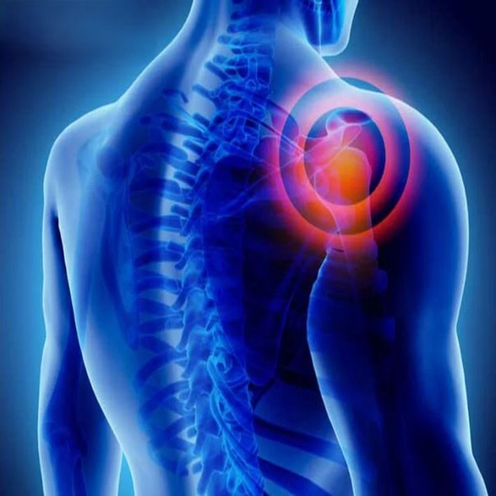
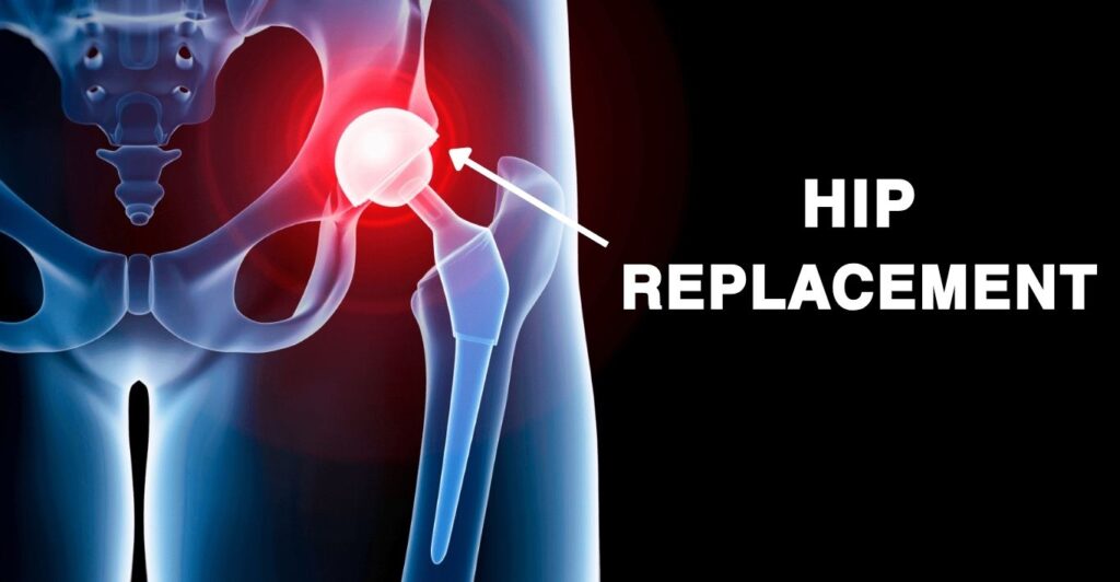
WHAT ARE SHOULDER FRACTURES?

Our shoulders are one of the most flexible and important joints in the body, enabling a wide range of movements. However, because of their mobility and structure, they are also vulnerable to injuries, especially fractures. A shoulder fracture occurs when there’s a break in one of the bones that make up the shoulder joint.

◼️Acromioclavicular Joint (AC Joint):

Located at the top of the shoulder, formed by the acromion (part of the scapula) and the clavicle.
◼️Glenohumeral Joint:
A ball-and-socket joint formed by the humeral head (“the ball”) and the glenoid (part of the scapula).

When any of these bones break, the result is a shoulder fracture, which can vary in severity and location.

TYPES OF SHOULDER FRACTURES?

Now you know that three bones form the shoulder joint; hence, the location of the break defines the type of shoulder fracture. Here are the three types you should know:

◼️Clavicle Fracture
A break in the collarbone, usually caused by trauma such as falls, motor vehicle accidents, or sports injuries.
◼️Proximal Humerus Fracture
This occurs in the upper part of the arm bone, near the shoulder joint. It often results from direct trauma, falls, or collisions. The humeral head, known as “the ball” of the joint, is typically involved.
◼️Scapula Fracture
The scapula (shoulder blade) is a strong bone, so breaking it usually requires significant trauma, such as high-impact sports injuries or automobile accidents.

SYMPTOMS OF SHOULDER FRACTURES?

Fractures are painful injuries, and shoulder fractures are no exception. Common signs and symptoms include:

◼️Severe shoulder pain
◼️Difficulty or inability to move the affected arm
◼️Swelling and bruising around the shoulder
◼️Tenderness to touch
◼️A visible bump at the fracture site (common in clavicle fractures)
◼️Discoloration around the upper arm

WHAT CAUSES SHOULDER FRACTURES?

◼️Automobile accidents: High-energy collisions are a leading cause.
◼️Sports injuries: Contact sports like football and hockey increase the risk.
◼️Falls: The direction of the fall often determines which part of the shoulder is injured.
◼️Direct trauma: A heavy blow or strike to the shoulder can also cause fractures.

For more information, talk to a healthcare provider.

If you have any questions about Shoulder Fractures, Please feel free to leave a comment.

Do share this blog with your friends and family!

Read More

WHAT IS OSTEOPOROSIS?

Osteoporosis is a bone disease where you suffer from excessive breaking of your bones, lesser creation of new bones, or both. This may result in the weakening of bones.

Osteoporosis means ”porous bone”. If you watch a healthy bone under a microscope, you will see a honeycomb. When you have osteoporosis, the holes and the spaces in the honeycomb are much larger than what you will see in healthy bones.

Osteoporosis affects both men and women. Women are more prone to Osteoporosis, especially women after menopause.

SYMPTOMS OF OSTEOPOROSIS?

In the initial stage, there are no signs or symptoms. But as Osteoporosis starts weakening your bones, you might start seeing the following symptoms.

◼️You start suffering from back pain, possibly caused by the fractured bone.
◼️You will begin to lose height over
time.
◼️You will have a stooped posture
due to the bending of your spine.
◼️You will see the breaking of bones more often.

(We recommend that you consult the Orthopedic doctor or visit an Orthopedic Hospital near you if you are facing severe issues)

STAGES OF OSTEOPOROSIS?

◼️Stage 1 (Age 25–30): Peak bone density. Bone formation equals bone loss. No visible
symptoms.
◼️Stage 2 (Age 30–35): Bone loss begins to exceed formation. Early signs may appear
as receding gums, brittle nails, and a weak grip.
◼️Stage 3 (Age 45–55): Noticeable bone fragility, especially in post-menopausal
women. Symptoms include fractures from minor falls, back or neck pain.
◼️Stage 4: Severe bone loss with deformities and height loss. Daily activities like
climbing stairs or sitting become painful and difficult.

CAUSES OF OSTEOPOROSIS?

◼️Aging — the main risk factor
◼️More common in post menopausal women
◼️Metabolic abnormalities
◼️Low calcium intake or deficiency
◼️Reduced bone formation rate
◼️Low estrogen or androgen levels
◼️Genetic disorders
◼️Conditions like bone cancer or thyroid issues
◼️Long-term use of corticosteroids

For more information, talk to a healthcare provider.

If you have any questions about OSTEOPOROSIS, Please feel free to leave a comment.

Do share this blog with your friends and family!

Read More

The tibia, commonly known as the shinbone, is one of the most important bones in the human body. Located between the ankle and the knee, it carries most of our body’s weight and plays a crucial role in walking, running, and everyday movement. Because of its load-bearing function, the tibia is also one of the bones most prone to fractures.

ANATOMY OF THE TIBIA:

The tibia is the larger of the two bones in the leg, positioned between the knee and the ankle. It has a hollow canal filled with bone marrow and a strong outer layer known as the cortex, which provides strength.

Fisioterapia Sportiva Milano - Dott. Creperio Fisioterapista

◼️Tibial plateau – top part of the tibia near the knee
◼️Tibial plafond – bottom part of the bone near the ankle.
◼️Tibial shaft – the central part where most of the fractures occur.

WHAT IS THE TIBIAL SHAFT?

The tibial shaft is the middle portion of the tibia, lying between the flared ends just below the knee (tibial plateau) and just above the ankle (tibial plafond). Unlike the ends, which are broader and flatter, the shaft is more triangular in shape.

SIGNS OF TIBIAL SHAFT FRACTURES:

Tibia / Fibula Fracture Specialist | 🩺Singapore Sports & Orthopaedic  Surgeon

Fractures of the tibial shaft typically occur with substantial traumatic injuries. Common signs of these fractures include:

◼️Sharp pain in the shin.
◼️Visible deformity of the leg.
◼️Bruising and swelling around the shin.
◼️Inability to bear weight on the leg.

Because such injuries often occur with other trauma, doctors usually examine the entire body to rule out
hidden injuries.

For more information, talk to a healthcare provider.

If you have any questions about TIBIAL SHAFT FRACTURES, Please feel free to leave a comment.

Do share this blog with your friends and family!

Read More

Causes of Ankle Fracture

The most common bone and joint injuries are ankle injuries. How you should be careful about your ankle injury is depending upon the degree of pain, inability to walk and if the bone is broken or not. Most of the time, the doctor also has the same concern, is there a broken bone? Because diagnosing sprain, dislocation or tendon injury is much easier than diagnosing a fracture.

Let’s see what the main causes of an ankle fracture are. But before that, we need to know the structure of the ankle joint. It is made up of 3 bones coming together.

The tibia- It is the main bone of the lower leg, makes up the inside of the ankle joint.

The fibula- It is a smaller bone that parallels the tibia in the lower leg. It makes up the outside of the ankle joint.

The talus- Both the tibia and fibula are known as the singular is malleolus. Together they form an arch that sits on top of the talus.

These 3 bones- the tibia, fibula, and talus make up the bony elements of the ankle joint. It is stabilized by several ligaments that hold these bones in place.

Ankle Fracture Causes

Stress is the most common cause of an ankle fracture. If you stress an ankle joint beyond strength, you injure the joint. If there is only wear and tear on the ligaments then you have sprained the ankle. In case of breaking bones, you have an ankle fracture. It can occur with simultaneous tears of the ligaments. These are the reasons for ankle fracture:

  • Twisting the ankle side to side
  • Rolling the ankle in or out
  • A strong force applied to the joint by coming straight down on it as in jumping from a high level
  • Flexing or extending the joint

This is the general information about the causes of Ankle Fracture. We recommend you to take your doctor’s advice for proper guidance. We hope this information adds value to your knowledge. Watch out this space for more such information. Greetings for SYS Medtech International PVT. LTD.

Read More

Types of tibia fracture

The long bone located in the lower leg between the knee and foot is known as the shinbone or tibia. Tibia fractures are usually caused by an injury or repetitive strain on the bone and it’s very common. The severity and type of fracture may vary, depending on the cause of the broken bone. Fractures that occur at the upper part of the tibia are called proximal fractures and fractures that occur below this area known as tibia shaft fractures. Apart from this, the following are the major types of tibia fractures-

  • Spiral fracture- A spiral-shaped fracture occurs when a twisting movement causes a break.
  • Displaced fracture- When a crack in the bone moves part of the bone so that it is no longer aligned, a displaced fracture occurs. Surgery is often needed to correct this type of fracture for realigning the bones back together.
  • Stable fracture- It involves a crack in the bone that leaves most of the bone intact and in its normal position. During the healing process, the broken parts of the tibia line up and maintain their correct position. This is also known as a non-displaced fracture.
  • Stress fracture- Stress fractures commonly known as hairline fractures are overuse injuries. These fractures are small as there are just thin cracks in the bone.
  • Comminuted fracture – When the bone fractures into three or more pieces, this is called a comminuted fracture.

When bones are broken, they can either stay under the skin or break through its surface. When a broken bone breaks through the skin, it is known as open fractures. With closed fractures, the bone does not break the skin, still, there may still be internal tissue damage.

This is the general information about Tibia Fracture. We recommend you to take your doctor’s advice for proper guidance. We hope this information adds value to your knowledge. Watch out this space for more such information. Greetings for SYS Medtech International PVT. LTD.

Read More

Femur fracture- Symptoms and causes

The bone located in the human thigh is known as the femur bone. It is the longest bone, extending from the hip to the knee. Also, it is the strongest bone in the human body. When the femur bone breaks, it takes a long time to heal. Breaking this bone can make everyday tasks much more difficult because it’s one of the main bones responsible for walking.

Symptoms of a broken femur

  • Immediate, severe pain
  • The injured leg appears to be shorter than the unbroken leg
  • Not able to put weight on the injured leg
  • The injured leg appears to be crooked

Causes of femur fracture

A femur fracture is very rare as it is a very large and strong bone. A broken femur is usually caused by a severe accident such as vehicle accidents. A break in this bone can only result from a large amount of force such as a fall from an extreme height. This fracture can take three to six months to heal.

Older adults have weaker bones. Thus they have the tendency of having a femur fracture. It may be called a hip fracture instead of a femur fracture, depending on how close the break to the hip.

A broken femur can have a major effect on your life, but not permanent. In most cases of broken femurs, patients return to a normal lifestyle. Surgeries become effective and people are typically able to completely heal from a broken femur.

This is the general information about Femur Fracture. We recommend you to take your doctor’s advice for proper guidance. We hope this information adds value to your knowledge. Watch out this space for more such information. Greetings for SYS Medtech International PVT. LTD.

Read More

Types of Shoulder Fractures

The scapula (shoulder blade), clavicle (collarbone) and humerus (upper arm bone) are three bones involved in shoulder fractures. All three types of shoulder fractures can cause shoulder pain, swelling and tenderness. Also, it limits the shoulder’s range of motion. Let’s see these three major types of shoulder fractures and how it affects the human body.

Scapula Fracture

A flat triangular bone located in the upper back is known as the scapula. The scapula provides the primary connections between the arm and the chest. Scapula fractures are rare which approximately less than 1% of all fractures are. A scapular fracture may result from a contact sport like football and cricket or a traumatic event such as a car or bike accident.

Clavicle Fracture

The clavicle, commonly known as collarbone is a long and thin bone that begins at the base of the neck and extends out to the shoulder. This fracture can happen at any age, from new-borns to the elderly. Normally, this fracture occurs because of a fall, direct hit or a motor vehicle accident. Common symptoms are bruising, swelling, and pain over the clavicle region.

Proximal Humerus Fracture

The humerus is that long bone between the elbow and the shoulder. A fracture to the top of the humerus bone is called a proximal humerus fracture. The ball that rests in the shoulder’s socket is often referred to as the humeral head. A proximal humerus fracture is very common and it can occur at any age. It can often be treated without surgery. Whether the patient needs the surgery is depending on various factors like the amount of displacement, the involvement of the cartilage in the joint, the age and activity level of the patient.

This is the general information about Shoulder Fracture. We recommend you to take your doctor’s advice for proper guidance. We hope this information adds value to your knowledge. Watch out this space for more such information. Greetings for SYS Medtech International PVT. LTD.

Read More

Dislocated Elbow- Symptoms and Causes

The elbow is made up of three bones- humerus, radius and ulna. The elbow dislocation happens when any of these bones become separated or knocked out of place. If a person thinks that he/she has a dislocated elbow, he/she should get immediate medical help because dislocation can be very painful, causing the elbow to become unstable and sometimes unable to move. It damages the ligaments of the elbow and can also damage the surrounding muscles, nerves and tendons.

Causes of a dislocated elbow

  • When people try to stop a fall with an outstretched hand
  • Car accidents
  • Sports injuries
  • A joint disorder such as Ehlers-Danlos syndrome makes joints unusually loose and flexible

Symptoms of a dislocated elbow

A dislocated elbow can be partial or complete. A complete elbow dislocation involves a total separation called a luxation and when the elbow joint is partially dislocated, it is called a subluxation. Doctors also classify elbow dislocations according to where it occurs and the extent of the damage. The 3 types are:

Simple: No major injury to the bone

Complex: Severe injuries to the bone and ligament

Severe: Damage to the blood vessels and nerves around the elbow

The symptoms of a dislocated elbow vary depending on the type of injury and the bones involved. These are the common symptoms-

  • Bruising
  • Deformed-looking arm (bone looks out of place)
  • Weakness in the joint
  • Loss of ability to move the elbow
  • Pain
  • Swelling

This is the general information about Elbow Fracture. We recommend you to take your doctor’s advice for proper guidance. We hope this information adds value to your knowledge. Watch out this space for more such information. Greetings for SYS Medtech International PVT. LTD.

Read More

Symptoms of Wrist Fracture

Breaking one or more bones of the wrist can cause a wrist fracture. A broken wrist occurs when people try to catch themselves during a fall and land hard on an outstretched hand. If you participate in sports like cricket or kabaddi, you may be at higher risk of a broken wrist. Also, if you have a condition in which bones such as osteoporosis where bones become thinner and more fragile, the chances of wrist fracture are high.

For bones to heal in proper alignment, it’s important to treat a broken wrist as soon as possible.  The delay in this might affect your ability to do everyday activities, such as writing, buttoning a shirt or knotting shoelaces. Also, early treatment helps in minimizing pain and stiffness. Thus one has to know the primary symptoms of wrist fracture so that the person can rush to the doctor.

Following are the symptoms of the broken wrist

  • Swelling
  • Severe pain while gripping or squeezing or moving hand or wrist
  • Bruising
  • Tenderness
  • Obvious deformity, such as a bent wrist

When to call a doctor

Looking at these symptoms, if you think you might have a broken wrist see a doctor immediately. If you’re having numbness, swelling or trouble moving your fingers you should rush to the doctor. A delay in diagnosis and treatment can lead to poor healing, restricts the motion and decreased grip strength.

This is the general information about Wrist Fracture. We recommend you to take your doctor’s advice for proper guidance. We hope this information adds value to your knowledge. Watch out this space for more such information. Greetings for SYS Medtech International PVT. LTD.

Read More

Why athletes should consider PRP treatment?

Many athletes face orthopaedic injuries often in their life which involve invasive procedures, complicated surgeries and lengthy hospital stays. This procedure usually needs a long period of time for recovery. Today with the help of technology and advanced medical science it is possible to undergo a variety of orthopaedic procedures using minimally invasive techniques such as PRP treatment.  So the athlete can get back to the active lifestyle more quickly than ever.

You must have read our previous blogs on PRP. If not then check them out on our website https://www.sysmedtechint.com/. In this blog, let’s see why you should opt for the PRP treatment.

To kick start your body’s healing abilities

Platelet-rich plasma or PRP is an ideal method of boosting your body’s own healing abilities. To simplify this, let’s take an example. Just think of the last time you cut your finger or scraped your leg enough to draw blood. When something like this happens, your body’s innate healing proteins are found in the platelets in your blood. This element rushes to the cut or scrapes to initiate the healing of your wound. PRP treatment boosts this ability.

A simple procedure

As you know the procedure of PRP treatment. There is no surgery and long stays in the hospital. You can go home after a few hours of this PRP treatment. You can resume your routine after a few weeks but under your doctor’s instructions.

Speedy recovery

As PRP injections send a high concentration of proteins directly to the injured area, these growth factors fasten up your body’s natural healing process.

Less risky

If you’re worried about taking medications or injecting artificial and manufactured fluids into your body then PRP is a more natural approach. It comes from your own blood, so the risk of rejection is highly unlikely. Thus there’s almost no risk of side effects. Besides minor irritation or pain at the site of the PRP injection, platelet-rich plasma therapy is one of the safest ways to heal your body after an orthopaedic injury.

This is the general information about PRP Treatment. We recommend you to take your doctor’s advice for proper guidance. We hope this information adds value to your knowledge. Watch out this space for more such information. Greetings for SYS Medtech International PVT. LTD.

Read More