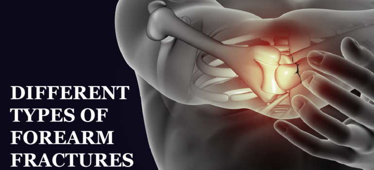
Different types of forearm fractures

The forearm is made up of a combination of two bones- the ulna and the radius. If a fracture happens in any one of the bones then it is known as a forearm fracture. Falling on the forearm or outstretched arm and direct impact from an object to the forearm are some of the causes of this type of fracture.

What is a forearm fracture?
As mentioned above, the forearm consists of two bones- the radius and the ulna, where the ulna is located on the little finger and the radius on your thumb side. Forearm fracture can occur at different levels. A distal fracture, which is near the wrist at the one end of the bone, in the middle of the forearm and a proximal fracture which is near the elbow at the top end of the bone. It can occur through a direct fall on the forearm or direct impact from an object or indirect injury. Landing on an outstretched arm is the secondary cause.


Different types of forearm fractures
Forearm fracture can either occur radius or ulna as a single fracture or a combination of both fractures. There are two types of forearm fracture- Galeazzi and Monteggia. The type of fracture depends on whether both bones are fractured at different levels or is there a joint injury at the wrist or elbow.
Galeazzi fracture: When a displaced fracture in the radius and a dislocation of the ulna at the wrist, where the radius and ulna come together, Galeazzi Fracture happens.
Monteggia fracture: When a fracture in the ulna and the top of the radius is dislocated at the elbow joint, called Monteggia Fracture.

This is the basic information about Forearm Fracture which is commonly known as Ulna Fracture. We recommend you to visit a doctor after facing such issues. We hope this information adds value to your knowledge. Watch out this space for more such information. Greetings for SYS Medtech International PVT. LTD.

Read More

What is Humerus Fracture?

The long bone of your upper arm is known as the Humerus. It extends from the shoulder to the elbow and it joins with the ulna and radius bones. Any break in this bone is referred to as a Humerus Fracture. A person may have either shoulder pain or elbow pain depending on where the fracture is and it takes several weeks for recovery.

Types of Humerus Fracture
Depending on the location of the crack, there are three types of Humerus Fracture-
Proximal- When there is a break in the upper part of the bone, it is called a proximal fracture. This causes shoulder pain as it is close to the shoulder.
Mid-shaft- This happens when there is a break in the middle of your humerus.
Distal- A distal humerus fracture occurs near the elbow. It is usually part of a more complex elbow injury and sometimes involves loose bone fragments.

What causes it?
Any Severe injury to the upper arm can result in a humerus fracture, but some are more likely to cause certain types. For example, falling with an outstretched arm can often cause mid-shaft and proximal humerus fractures. A high-impact crash, such as a car accident or football tackle, is more likely to cause a distal humerus fracture. If there is a medical condition where bones are becoming weaker, then humerus fracture may occur as a pathological fracture. This can lead to injuries and fractures from everyday activities that wouldn’t usually cause any injuries.

Things that can cause pathological humerus fracture-
Bone Cancer
Bone Infection
Osteoporosis
Bone Cysts or Tumors

This is the basic information about Humerus Fracture. We recommend you to visit a doctor after facing such issues. We hope this information adds value to your knowledge. Watch out this space for more such information. Greetings for SYS Medtech International PVT. LTD.

Read More

Causes of distal radius fracture

If a person gets hit or slips, he first puts his hands on the ground to balance himself. This often causes great strain on the wrist and can lead to fractures. This problem can happen to people of any age, but it is more common in older and younger children particularly older women.

There are reasons why the distal radius fracture happens:

For children about ages 8 to 14, there is a period during which bones have grown longer because of growth spurts. But the bone mineral density has not yet caught up which leaves the newly-grown bones more vulnerable to fractures. Chances of fracture get increased, particularly during activities such as sports or play.

In the case of senior citizens, their hormone levels shift and bone mineral density starts to decrease. This is especially happening with women once they reach menopause. Their risk of a distal radius fracture begins to rise at age 50 and that risk doubles every 10 years. For men, the related risk is much later in their life, at the age of 80.

Falling onto a wrist is the most common cause of a distal radius fracture, people who actively participate in sports that involve potential falls, such as Kabaddi, Hockey, Football or Cricket, are also at greater risk for this injury.

Also, car accidents or similar traumatic events can also cause a distal radius fracture. But the majority of these cases are the result of falls.

Read More

Diagnosis of hip fracture


Hip fracture is known to be the most common fracture. It is especially prevalent in older people. After age 65, human bones become weak and it almost requires surgery. This injury can often be life-threatening. Thus it needs to be treated quickly. Before taking any decision let’s get to know about how to diagnose the hip fracture.

During a physical exam, the doctor will ask for your complete medical history. Also, the physician will look for any injuries commonly associated with hip fractures. To determine whether an open fracture has occurred, the doctor will examine any lacerations and determine how stable the hip is will press on the front and back of the pelvic area. To look for haemorrhage that may signify bone penetration into the rectum, a rectal examination also may be performed.

Tests for a broken hip:-

X-ray

X-radiation or commonly known as X-ray uses invisible high energy electromagnetic radiation to produce images of internal tissues, bones and organs onto film. It is usually sufficient for the majority of fractures.

MRI (Magnetic Resonance Imaging)

It is a medical imaging technique that uses a combination of large magnets, radio frequencies and a computer. It is used to produce detailed images of organs and structures within the body; especially useful for assessing soft tissue around injured joints and bones.

Computed tomography scan (CT scan)

It uses a combination of x-rays and computer technology to produce 3D cross-sectional images, both horizontally and vertically, of the affected area, including bones, fat, soft tissue.

Bone scan

It uses a radioactive dye to visualize the bones. It’s different from plain x-rays or CT in that because of a nuclear medicine imaging technique, it shows bone metabolism and cell activity in the bones. Bone scans are used to assess including cancer of the bone or metastasis, location of bone inflammation and fractures, and bone infection.

This is how one can diagnose a hip fracture. We recommend you to visit a doctor instead of doing it by yourself. We hope this information adds value to your knowledge. Watch out this space for more such information. Greetings for SYS Medtech International PVT. LTD.

Read More

Anatomy of the Spine

When encouraging or applauding someone, we always say keep it up. The spinal cord is the most important part of the human body. In fact, it is safe to say that the human body depends on this single particle. But as mentioned above, is it really possible for a person to live with a stiff spine? Let’s take a closer look at this blog.

The human spine is made up of three segments. These segments form three natural curves when viewed from the side. The c-shaped curves of the neck and the lower back, which is called the cervical spine and the lumbar spine respectively, and the reverse c-shaped curve of the chest is called the thoracic spine. Let’s understand the structure of the spine-


Vertebrae
The human spine is made up of small bones known as vertebrae. These are the bones, which are assembled on top of one another which creates the natural curves. The spinal cord and nerve roots get protected by these bones as they create a canal. There is a total of 7 cervical vertebrae, 12 thoracic vertebrae, and 5 lumbar vertebrae.

Spinal Cord and Nerves
The spinal cord travels through the middle part of each stacked vertebra, called the central canal which extends from the skull to your lower back. Nerves carry messages between the brain and muscles by branching out from the spinal cord through openings in the vertebrae.

Muscles and Ligaments
Muscles work to stabilize and support the spine. Also, the ligament keeps the spine in good shape and helps in coordination.

Intervertebral Disks
These sit in between the vertebrae. They are flat and round. These made up of two main components- Nucleus pulposus and Annulus fibrosus. These disks act as shock absorbers for the spine as they maintain the strength of the spine by expanding when required.

Facet Joints
There are small joints between the back of the vertebrae that help the human spine move. There are four facet joints associated with each vertebra. The facet joints are important as they allow rotation of the spine.

This is the anatomy of the spine. I hope this information adds value to your knowledge. Watch out this space for more such information. Greetings for SYS Medtech International PVT. LTD.

Read More

Overview of Skeleton

The human skeleton is the basic structure of the human bones that holds the human body. The human skeleton is made up of many different types of bones and cartilage. Fibrous tissues called ligaments and tendons are also part of the skeleton. In this blog, we are going to get information about these bones and their structure.

Like other vertebrate species, the human skeleton has two main subdivisions, these are- axial and appendix. The axial includes the vertebral column, which is the spine and most of the skull. The appendix consists of the pelvic and pectoral lumbar and cartilage of bones. The third subdivision is visceral which includes the lower jaw, the upper jaw, and branched arches with the hyoid bone. Each subdivision has specific characteristics.

The human skeleton has three main functions- support, protection and motion. Of these functions, support is the most primary and basic function of the skeleton which might be the reason for its existence. All internal organs, systems and muscles depend on skeletal support.

After that, the human skeleton protects the internal organ from severe damage. For eg. the rib cage protects the lungs and heart, the skull protects the brain, etc. Looking at these examples we can surely say that the development of the human skeleton is the most important part of the evolution of mankind.

The third common function of the skeleton is the motion. Every muscle in the body is linked to the skeleton. Thus, from a beautiful straight drive by Sachin Tendulkar to the strokes on canvas by an artist and from the adventurous expeditions made by ISRO astronauts to the potter who works hard to make pottery, the movements of the body and its organs have been made possible by independent engineering systems of muscles and bones.

This is the overview of the human skeleton. I hope this information adds value to your knowledge. Watch out for more knowledge. Greetings for SYS Medtech International PVT. LTD.

Read More